
Most breast imaging is designed to answer one question: Is something there?
A mammogram looks for masses and calcifications. An ultrasound helps determine whether a lump is solid or filled with fluid. These tests show structure: what the tissue looks like and what’s inside it.
Thermography asks a different question: How is the tissue behaving?
Breast tissue is always active. Blood vessels widen and narrow. Hormones influence growth and sensitivity. Cells use energy and respond to signals from throughout the body. Much of this happens quietly, without pain, a lump, or any change you’d notice.
Thermography offers one way to observe patterns associated with that activity.
What Is Breast Thermography?
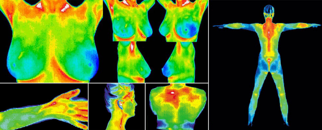
Every part of your body gives off heat in the form of infrared energy. Thermography uses a specialized camera to detect that energy and turn it into a color map of your skin’s surface temperature. Different colors represent warmer and cooler areas.
The exam involves no compression, physical contact, or ionizing radiation.
Think of a mammogram as looking at a house’s framing. It shows the walls, the beams, and anything that doesn’t belong. Thermography is more like pointing a thermal camera at the outside of the house. It can show which areas are running warm, but not what’s causing it.
Where Does That Heat Come From?
Heat is a natural byproduct of cells doing their work. Blood carries that heat toward the skin, and small blood vessels widen or narrow depending on what the body needs.
Several things influence the heat patterns across your breasts:
- Circulation: Blood flow affects how heat is distributed through the tissue.
- Hormones: Normal hormonal shifts can influence breast tissue and blood vessels.
- Inflammation: Inflammation can increase local blood flow and warmth.
- The nervous system: Your nervous system helps regulate blood vessels in response to temperature and other signals.
- Your individual anatomy: Breast composition, skin characteristics, and blood vessel patterns differ from woman to woman.
These influences overlap. A warmer area may have several possible explanations, and a thermogram records their combined effect.
What Thermography Can and Can’t Tell You
Different imaging tools answer different questions.
Mammography uses low-dose X-rays to look at breast structures, including masses and calcifications.
Ultrasound uses sound waves to examine tissue and can help distinguish fluid-filled cysts from solid findings.
Breast MRI uses magnetic fields, usually with contrast dye, to create detailed images of breast tissue.
Thermography shows surface temperature patterns influenced by physiological activity.
This difference is what makes thermography interesting. Like the thermal camera pointed at the house, it may reveal an unusual pattern without identifying what’s causing it beneath the surface.
Thermography cannot diagnose breast cancer, so it is used alongside recommended mammography and other appropriate screening, rather than in place of them.
What Does the Research Say?
Researchers have studied thermography for decades, exploring whether changes in blood flow and tissue activity can sometimes appear before a structural abnormality becomes visible on imaging.
Newer research has brought fresh attention to the technology.
A 2024 analysis combining 22 studies found that thermography identified approximately 9 out of 10 cancers in the populations studied. It also flagged roughly 3 out of 10 people who did not have cancer.
The findings suggest that thermography can recognize thermal differences associated with breast abnormalities, while interpreting what those differences mean remains more challenging.
Researchers are continuing to investigate improved cameras, computer-assisted interpretation, and methods for measuring how tissue responds to changes in temperature.
An important question is which patterns indicate meaningful physiological changes and which reflect normal variation.
Why a Baseline Matters
No two women have exactly the same breast tissue. A first thermogram can provide a reference point for future comparisons.
When you return for another scan under similar conditions, the images can be compared for changes in symmetry, temperature distribution, and visible vascular patterns. Knowing whether a pattern has remained stable or changed gives the findings additional context.
Because the camera measures skin temperature, consistency matters. At a typical appointment, you’ll review your health history and spend some time in a temperature-controlled room while your skin adjusts. A technician then captures images from several angles so both breasts can be compared.
Some appointments include a brief cooling challenge followed by additional images. Known as dynamic thermography, this allows changes in surface temperature following a cooling stimulus to be observed.
A baseline gives you a starting point for following changes over time.
What Do the Ratings Mean?
Thermography results are often described using a rating scale from TH1 to TH5.
| Rating | What it describes |
| TH1 | Symmetrical, with little visible vascular activity |
| TH2 | Symmetrical, with visible vascular activity |
| TH3 | Unclear or questionable findings |
| TH4 | Abnormal findings |
| TH5 | Markedly abnormal findings |
These ratings describe the appearance of thermal patterns. They are not diagnoses or measurements of disease severity.
A TH3 or higher rating may lead to further evaluation, such as an ultrasound or MRI, depending on your history, symptoms, examination, and previous results.
One Tool in a Larger Picture
Your breasts respond to changes throughout your body. Blood flow, hormones, and tissue composition can all shift over time.
Thermography records one aspect of that activity. Its findings can be considered alongside your health history, physical examination, symptoms, and other appropriate testing.
Understanding what each assessment measures can help you make more informed decisions about your breast care.
Related Articles
Looking Deeper
Most changes in the body develop gradually. Tissues adapt to shifting hormones, circulation, inflammation, stress, and other influences. Sometimes these changes begin well before symptoms appear.
This is a central idea in Foundational Medicine. Health and disease develop through biological processes that unfold over time.
Breast tissue participates in those same processes. Understanding how it functions adds another perspective to what structural imaging can show.
No single test captures every aspect of breast health. Following changes over time, recognizing what’s normal for your body, and knowing when to investigate something new all contribute to lifelong breast care.
Continue Your Discovery
Thermography is one way to learn about your breast tissue. Understanding breast health means considering the whole picture, from structure and hormones to circulation and lymphatic flow.
Denali’s Best Breast Health approach considers breast tissue and the biological environment surrounding it. The program includes establishing a personal breast-health baseline and individualized, hands-on instruction in lymphatic breast care, helping women take an active role in caring for their breasts throughout life.
Discovery Before Direction.
FAQ
What does breast thermography measure?
Breast thermography measures infrared energy emitted from the skin, creating an image of surface temperature patterns. These patterns are influenced by circulation, temperature regulation, and other physiological processes.
Can thermography detect changes before a lump forms?
Some research has investigated whether unusual temperature patterns can appear before structural abnormalities become apparent. However, thermography cannot reliably predict whether or when breast cancer will develop.
What does a TH3 thermography rating mean?
TH3 generally describes questionable or equivocal thermal findings. Depending on a woman’s history, symptoms, and other findings, further evaluation with ultrasound or another imaging method may be recommended.
Why is a baseline thermogram useful?
A baseline provides an initial record of breast temperature patterns. Future examinations performed under comparable conditions can be evaluated for changes in symmetry, temperature distribution, and vascular patterns.
Does breast thermography replace mammography?
No. Thermography measures surface temperature patterns, while mammography examines internal breast structures. Thermography does not replace recommended mammography or other appropriate breast-cancer screening.